
Figure 3.
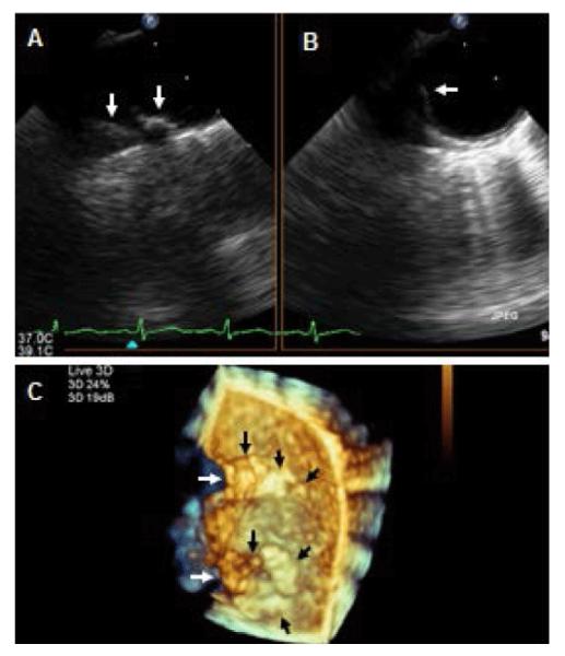
Two-dimensional and three-dimensional TEE for assessment of aortic atherosclerosis. A,B: Biplane 2-D TEE views of the distal arch and proximal descending thoracic aorta in a patient with SLE demonstrating a complex and protruding atherosclerotic disease located on the anterior and medial portions of the aorta (arrows) C: Three-dimensional en-face view of the anterior and in part medial and lateral walls of the distal portion of the aortic arch and proximal descending thoracic aorta demonstrating a tortuous aortic medial wall (white arrows), protruding atherosclerosis on the medial wall of the distal arch and proximal descending thoracic aorta (black arrows), and multiple other early atherosclerotic plaques in both aortic portions (short black arrows).