
Figure 4.
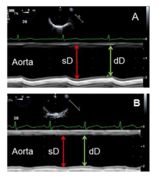
Assessment of aortic stiffness. A: Two-dimensional guided M-mode images of the mid descending thoracic aorta in a 42 year old SLE patient with normal aortic distensibility based on significant increase in aortic diameter during systole (sD) as compared to diastole (dD). B: Two-dimensional guided M-mode images of the mid descending thoracic aorta in a 39 year old SLE patient with aortic stiffness based on minimal change in aortic diameters during systole and diastole.