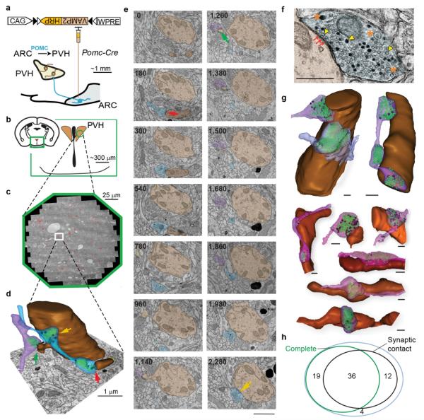
Figure 2.
GESEM-labeling for identification and reconstruction of ARCPOMC→PVH circuits. a, Schematic drawing for targeting Cre-dependent rAAV to POMC neurons for VAMP2:HRP expression b,c, Location (b) and aligned electron micrographs (c) of imaged PVH area from a mouse brain expressing VAMP2:HRP in POMC neurons (red: DAB-labeled boutons from one POMC-Cre mouse). d, Two reconstructed ARCPOMC→PVH axon segments (purple and blue) (Green: SVs, Black: LVs) making synaptic contacts (arrows) with a dendrite and a spine in the PVH (brown). e, Multiple z-aligned electron micrographs for the reconstructed terminals in d (numbers at upper left indicates position in z-axis in nanometers, based on 60 nm section thickness). Arrows, synaptic contacts. Scale, 1 μm. f, Example electron micrograph illustrating a synaptic contact (arrows) as well as labeled and unlabeled SVs (arrowheads) and LVs (asterisks). Scale, 0.5 μm. g, Other example ARCPOMC→PVH bouton reconstructions. Green: SVs, Black: LVs. Scale, 0.5 μm. h, Venn diagram of all boutons containing DAB-labeled vesicles categorized for completeness within the imaged volume and the presence of a post-synaptic partner. Most labeled boutons resided completely within the imaged volume (77%), and the majority (68%) of the boutons from POMC neurons had ultrastructural characteristics of a synaptic contact.

