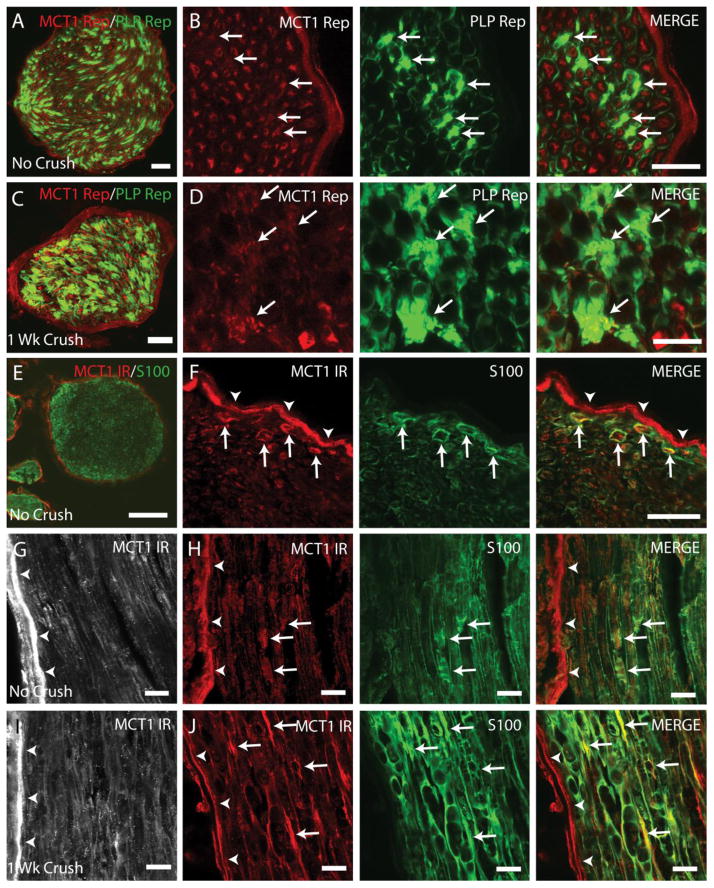
Figure 6. MCT1 is expressed by Schwann cells in intact and crushed peripheral nerves.
Cross-section of uncrushed (A, B) and 1 week crushed (C, D) sciatic nerves from MCT1 tdTomato and PLP-GFP double reporter transgenic mice (Scale bars A, C 50 μm; scale bars B, D 20 μm). Schwann cells expressing MCT1 (arrows) are indicated by co-localization of MCT1 reporter (MCT1 Rep; red) with PLP reporter (PLP Rep; green). Perineurium is indicated by arrowheads. Red fluorescence in MCT1 tdTomato reporter mice reflects mRNA gene expression within a given cell, but not specific transporter localization within the cell (i.e., dendrite versus axon or cell body). Uncrushed sciatic nerve from WT mice immunostained with MCT1 (red) and S100 (green) displayed in cross-section (E, F) and longitudinal (G, H) photomicrographs (Scale bar E 100 μm; scale bar F–J 20 μm). Sciatic nerves 1 week following crush (I, J) immunostained for MCT1 (red) and S100 (green). MCT1-immunreactive profiles that co-localize with S100 (i.e., Schwann cells) are indicated by arrows and perineurium by arrowheads.

