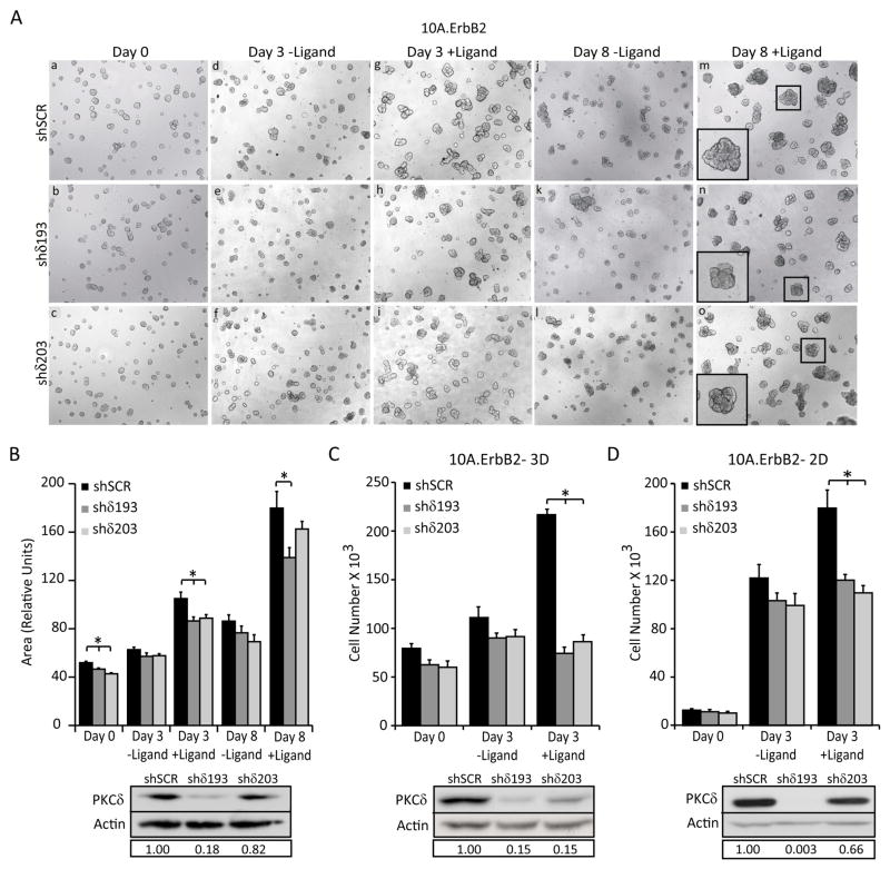
Figure 3. PKCδ is required for ErbB2-driven proliferation.
For all panels: PKCδ was depleted using lentiviral shRNA constructs (shδ193 and shδ203) and compared to control shRNA (shSCR) as described in Materials and Methods. A. 10A.ErbB2 cells depleted of PKCδ using shRNA (shδ193 and shδ203) were grown on Matrigel for 6 days (a, b, c). Cells were then left untreated (d, e, f; j, k, l) or treated with 1μM ligand for 3–8 days (g, h, i; m, n, o). Representative images of three separate experiments taken at 5X magnification are shown. Inset shows digital enlargement to show structure morphology. B. Quantification of structure area from Figure 2A, using Metamorph software. Representative of three experiments shown; *P=<0.02. D. and E. 10A.ErbB2 cells depleted of PKCδ were plated at equal densities in 3D (D) or 2D (E) growth assays. For 3D growth, cells were plated on Matrigel and grown for 6 days. Ligand was added for 3 days after which cells were tryspinized and counted; *P=<0.0001. For 2D growth, cells were treated with or without ligand for 4 days, trypsinized, and counted; *P=<0.04. B, C, and D. Insets show depletion of PKCδ by immunoblot with actin as a loading control. Boxed region below blots shows relative PKCδ knockdown compared to shSCR controls. Representative graphs of three separate experiments are shown.

