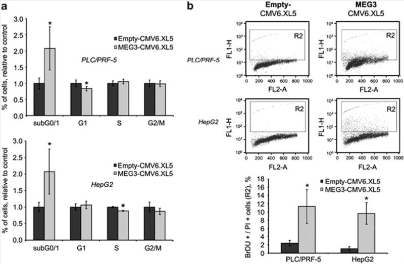
Figure 3.

MEG3 induces apoptosis. HepG2 and PLC/PRF/5 cells were serum-starved for 48 h and then transfected with 1 μg of MEG3-CMV6.XL5 or empty vector. After 24 h cells were collected and fixed in paraformaldehyde and ethanol overnight. (a) Cells were stained by propidium iodide (PI) and analyzed by flow cytometry with a BD FACScalibur (BD Bioscience, Heidelberg, Germany). Bars represent mean and standard error of three experiments expressed as relative to empty control. *P<0.05 compared with empty controls. (b) Apoptotic cells were detected by labeling DNA breaks using the terminal deoxynucleotide transferase dUTP nick end labeling assay (Invitrogen, Carlsbad, CA, USA). Cells were processed as indicated by the manufacturer, stained by PI and bromodeoxyuridine (BrdU), and analyzed by flow cytometry with a BD FACScalibur. Bars represent mean and standard errors of three independent experiments. *P<0.05 compared with empty controls.
