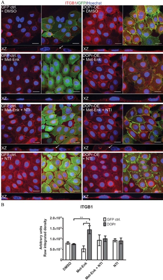
Figure 3.

δ-opioid receptor (DOPr) activation causes changes in β1 integrin (ITGB1) expression and distribution that can be partially reversed by δ-opioid receptor antagonists. (A) Confocal images of Met-Enk-treated GFP control and DOPr-OE N/TERT-1 cells labelled with antibodies recognizing GFP fluorescence tag (green), and ITGB1 (red), with Hoechst 33258 nuclear counterstain (blue). Orthogonal slicing of XZ-plane images is shown. Scale bar 20 μm. (B) Quantification of ITGB1 integrated intensity per image from average z-projected images. The results are expressed as means ± SEM of two independent experiments with two images taken per condition. Statistical comparison was done by one-way anova with Newman–Keuls post hoc test. *P ≤ 0.05 and **P ≤ 0.01.
