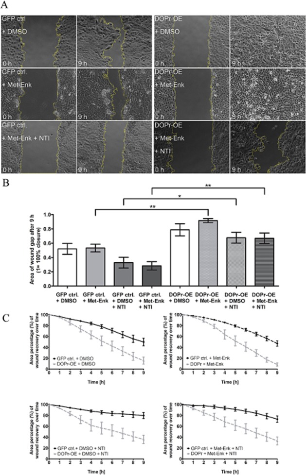
Figure 4.

Activation of δ-opioid receptors (DOPr) by Met-Enk enhances keratinocyte migration. (A) Time-lapse microscopy over 9 h of in vitro wound healing using GFP control and DOPr-OE N/TERT-1 cells. Panels show representative images of the keratinocyte monolayer at the gap, at 0 and 9 h post-wounding. Graphs depict quantified average area (B) of gap closure at 9 h (1 = 100% closure) and percentage (C) of gap closure (gap at time 0 = 100%) over time. Bar chart shows mean ± SEM (n = 11 fields of view from three independent experiments) for all groups (one-way anova, with Newman–Keuls multiple comparison test, *P ≤ 0.05 and **P ≤ 0.01).
