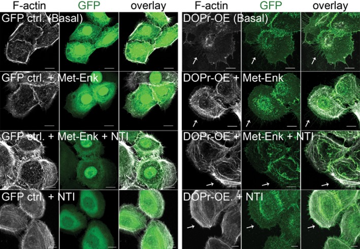
Figure 6.
Morphological changes in vitro in δ-opioid receptor (DOPr)-OE (overexpressing) N/TERT-1 keratinocytes. Confocal images of GFP control and DOPr-OE N/TERT-1 cells labelled with anti-GFP antibody (green) and Alexa 594-conjugated phalloidin (grey). GFP control N/-TERT-1 cells generally do not display fine protrusions while obvious fine protrusions containing F-actin at the rear of DOPr-OE N/TERT-1 cells were observed (white arrows). Fewer protrusions were observed when DOPr N/TERT-1 cells were treated with DOPr antagonist NTI. Scale bar 10 μm.

