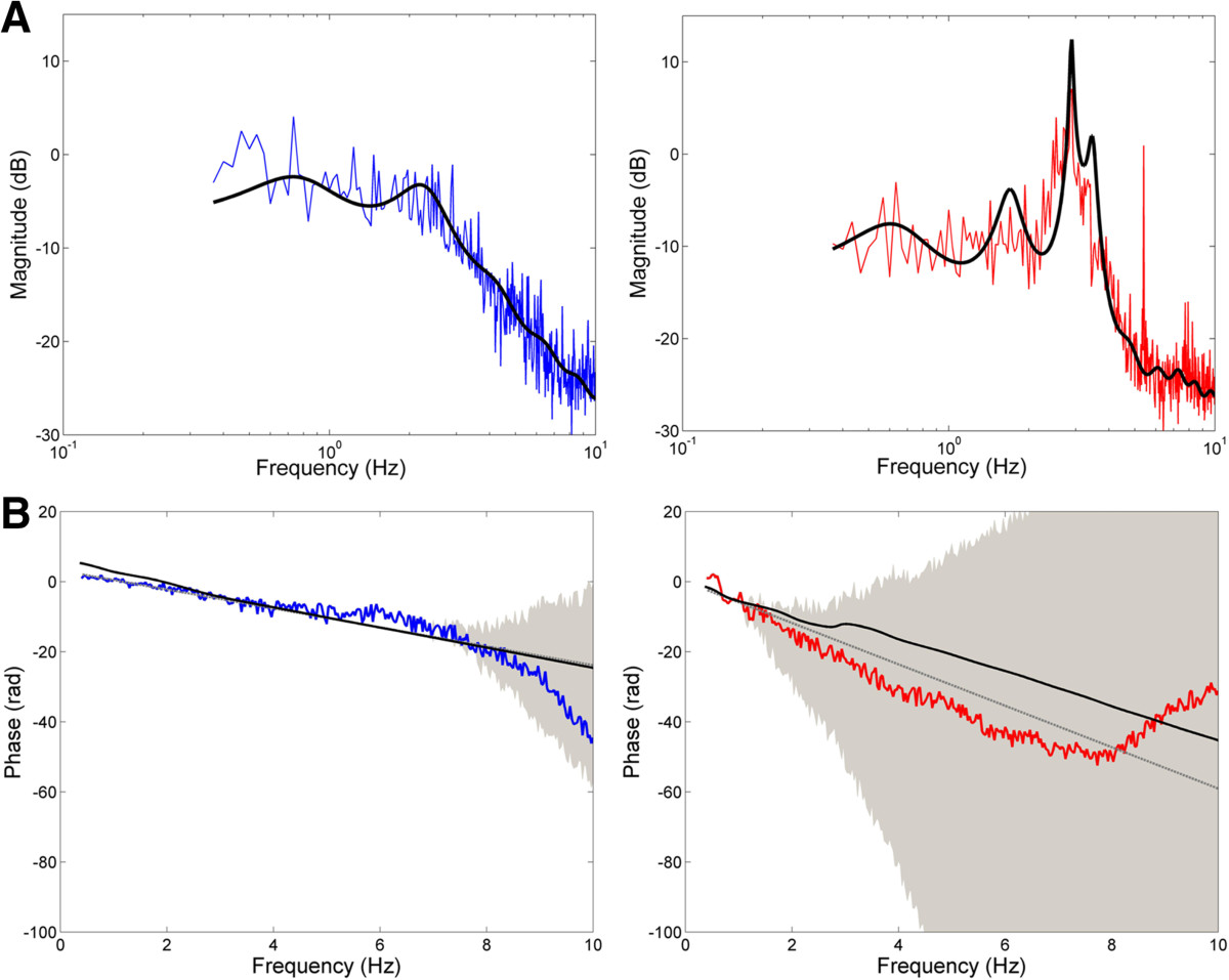
Figure 7.

Subject frequency response functions (FRFs) and model fits. (A) Magnitude of the FRF (colored traces) relating applied cursor perturbation to corrective change in arm position for subject 4 with MS (TAS = 2; right) and the age-matched control subject (left). The best-fit model for each subject is denoted by the solid black line. (B) Phase of the FRF (colored traces) with 95% confidence intervals (grey shading) for subject 4 with MS (right) and age-matched control subject (left). The solid black line denotes the best-fit model to the subject’s magnitude FRF. The grey line denotes the phase profile associated with the subject’s visual delay.
