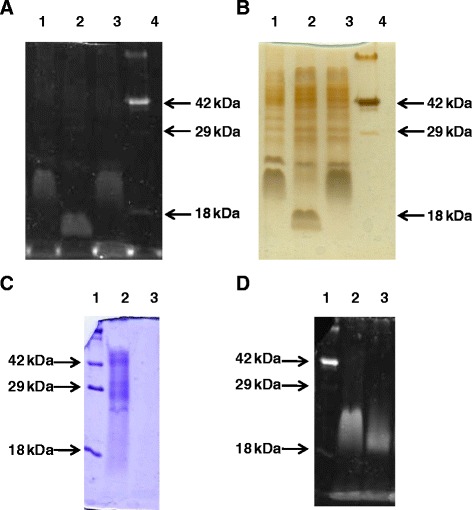
Figure 3.

SDS-PAGE and gel staining of glycosylated proteins extracted from F. psychrophilum wild-type, fpgA − , and fpgA + (complemented fpgA − ) strains. Total extracted proteins were separated by 14% SDS-PAGE gels that were (A, D) Pro-Q Emerald specific protein glycosylation; (B) silver; and (C) Coomassie blue, stained. (A) and (B): lane 1, parental; lane 2, fpgA-; and lane 3, fpgA+. In the right of each image, the position of Candy Cane (Life Technologies, Carlasbad, Cal.) with the 42 kDa and 18 kDa glycosylated proteins is indicated (lane 4). (C) and (D): lane 2, wild-type strain; lane 3, wild-type strain extract treated with proteinase K (similar results were obtained when trypsin and a mixture of both enzymes were used). Lane 1, Candy Cane (Life Technologies, Carlasbad, Cal.) molecular mass markers.
