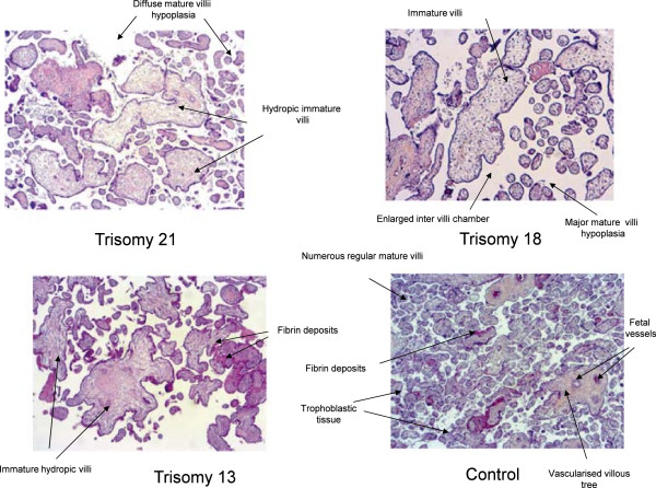
Figure 3.

Microscopic analysis of placental villi in normal pregnancy and pregnancy complicated by aneuploidy. After termination of pregnancy, 55 aneuploid placentae were analysed by experimented pathologists at the macroscopic level, and the histological level after being fixed in formalin, included in paraffin and stained with hematein-eosin-saffron. Mature villi i.e. small diameter, strong staining, presence of fetal vessels, decreased connective tissue, covered by trophoblastic tissue. Immature hydropic villi i.e. large diameter, pale staining, few fetal vessels, connective tissue, decreased trophoblastic tissue.
