
Figure 4.
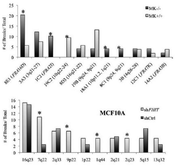
CFSs in epithelial cells with and without FHIT protein expression. Upper panel, comparison of frequencies of Aph-induced breaks at individual CFSs in the Fhit−/− and Fhit+/+ MK cells. Asterisks note fragile sites that vary extensively in frequency in the cells with absence of Fhit expression. The human homologous region for each mouse CFS is noted in parentheses. Lower panel, a similar comparison of numbers of breaks at CFSs in MCF10A cells infected with shCtrl or shFHIT lentivirus vectors; the order of frequency of the most fragile loci varies slightly in the two cell types, but the 16q23/WWOX locus remains the most frequently expressed in both cell lines.