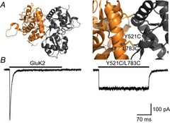
Figure 4. Crosslinking of the GluK2 LBD dimer interface yields non-decaying current responses.

A, side views of the LBD dimer interface of GluK2 Y521C/L783C (PDB 2I0C), in its entirety (left) and close-up (right), detailing the inter-protomer disulphide bond (yellow). B, activation profiles of wild-type GluK2 and Y521C/L783C receptors in response to 10 mm l-Glu (holding potential –60 mV). Adapted from Daniels et al. (2013) with permission.
