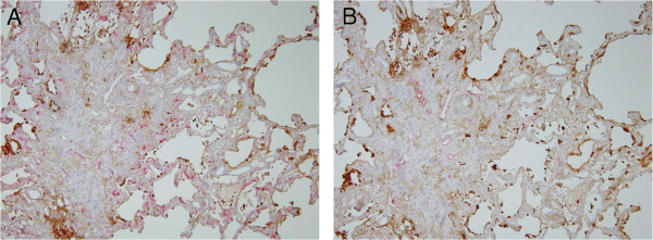
Figure 5.

Immunostaining of blood and lymphatic vessels in lung biopsies from idiopathic pulmonary fibrosis (IPF) patients. (A) The capillary vessels were extremely developed, especially along the air space (red: CD34, brown: SP-A). (B) On the other hand, lymphatic vessels were localized at the center of the stroma (red: D2-40, brown: SP-A).
