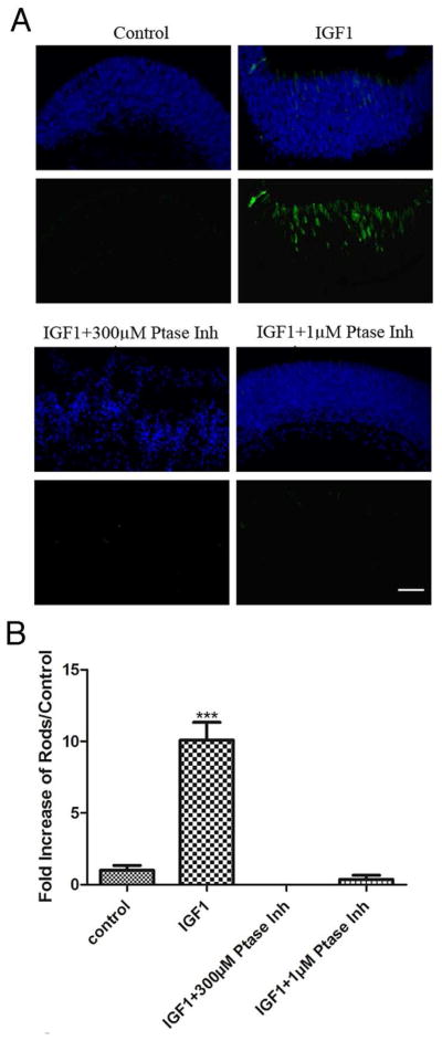
Figure 6. Inhibition of Shp1/2 during IGF1 treatment of retinal explants prevents IGF1-induced promotion of rod photoreceptor development.
(A) Immunofluorescence detection of opsin (green) overlaid with nuclear counterstain (Hoechst, blue) of four day P1 retinal explants. P1 retinas were cultured from WT mice for four days in the presence of 50ng/mL IGF1, or (B) Numbers of rods present on P1 retinas after four days of culture in the presence of 50ng/mL IGF1, 50ng/mL IGF1and 300μM NSC87877 and 50ng/mL IGF1 and 1μM NSC87877. Counts were obtained by averaging the number of rods present in three individual histological cross sections of each retina. At least three different retinas were studied per treatment focusing on the central areas of the retina. ***P<0.005. Scale bar in A, 40μm.

