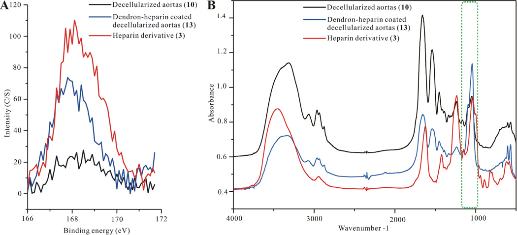
Figure 4.
A) XPS verification of heparin attachment on “click” coated decellularized aortas, B) FTIR verification of heparin attachment on “click” coated decellularized aortas. In both XPS and FTIR the modifications are color identified where the control untreated decellularized aortas are black; dendron coated decellularized aortas are blue; and dendron-heparin coated decellularized aortas are drawn in red.

