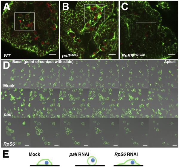
Figure 5. PALL promotes Actin remodeling during AC clearance.
(A-C) Confocal micrographs of Actin (green) and CRQ (red) immunostaining of yw embryos (wild-type (WT) control in A), pallko260 (B) and RpS6WG1288 (C) mutant embryos. (D) Confocal micrographs of Mock-treated, pall- or RpS6-RNAi S2 cells stained with phalloidin. Z-stack images through the cells were collected 1.74 μm apart (basal membrane is in contact with the glass slide). Scale bars correspond to 10 μm. (E) Schematic drawings summarize F-actin staining (green) distribution and cell shape for mock-treated, and pall- and RpS6-RNAi S2 cells seen in D. Green lines and blue circles represent Actin and nuclei, respectively. Black lines represent the slides. See also Fig. S4.

