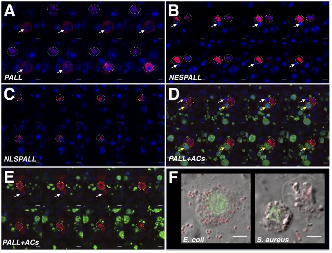
Figure 7. Regulation of PALL localization by ACs confers its specificity to efferocytosis.
(A-E) Z-stack confocal images through DRAQ5- (A-C) or DAPI- (D-E)(in blue) and HA Ab (in red) double-stained S2 cells expressing various forms of PALL. (A) A HA-PALLFL expressing S2 cell that has engulfed an endogenous AC (white arrow) shows both nuclear (dotted circles surround cell nuclei of interest) and cytoplasm HA-PALLFL protein expression, while a cell that has not engulfed nor bound to AC shows HA-PALLFL strictly in its nucleus. (B) HA-NESPALL expressing S2 cells where the L residues of the NES of PALL were mutated into A that prevents PALL nuclear export even when cells have engulfed endogenous ACs. (C) HA-NLSPALL expressing S2 cells where the NES was replaced by a NLS show its nuclear localization. (D-E) A HA-PALLFL expressing S2 cell that has engulfed an endogenous AC (white arrow) and a FITC-labeled AC (yellow arrow) (D), and a HA-PALLFL expressing S2 cell that is engulfing an endogenous AC (white arrow)(E) showing both nuclear (dotted circle around nucleus) and cytoplasmic localization of HA-PALLFL. (F) Nuclear localization of PALL on confocal micrographs of HA Ab-stained (green) HA-PALLFL-expressing S2 cells that have engulfed E. coli and S. aureus bacteria. Scale bars are 5 μm. See also Fig. S6.

