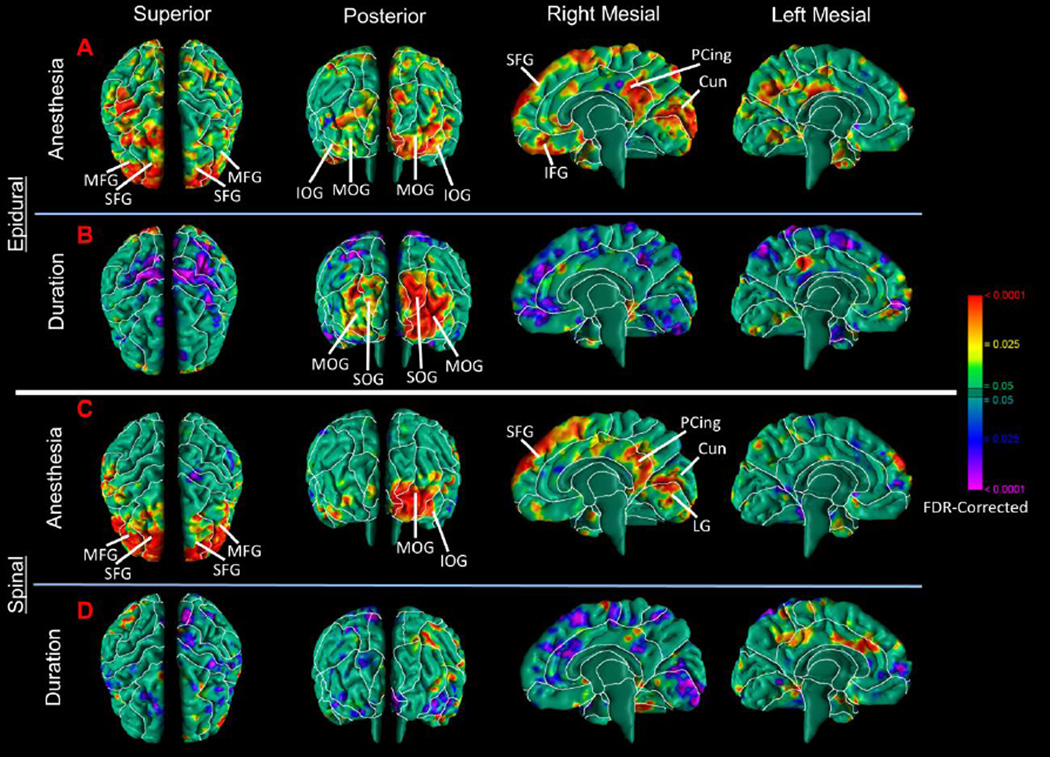
Figure 2. Correlation of the Duration of Anesthesia Administration with Local Volumes.
Maps are shown for differences in morphological measures of the cerebral surface in infants exposed to epidural or spinal anesthesia separately. (A) Epidural anesthesia-exposed infants demonstrated a significant increase in local volumes of the frontal lobes bilaterally and the occipital lobes predominantly on the right, and the posterior portion of the cingulate gyrus on the right hemisphere. These findings are similar to the main effect of regional anesthesia combined (Fig.1). (B) The duration of anesthesia exposure, an estimate of the dose that anesthesia-exposed infants received, correlated positively with local volumes in the occipital lobe bilaterally. (C) Spinal anesthesia-exposed infants had significant increases in local volumes of the frontal lobes bilaterally and occipital lobe on the left and the posterior portion of the cingulate gyrus on the right hemisphere. These findings are similar to the effect of regional anesthesia combined and epidural anesthesia. (D) The duration of anesthesia exposure, an estimate of the dose that anesthesia-exposed infants received, did not correlate significantly with local volumes in brain regions identified in the primary regional anesthesia analyses. Abbreviations: SFG, superior frontal gyrus; MFG, middle frontal gyrus; IFG, inferior frontal gyrus; PCing, posterior cingulate; SOG, superior occipital gyrus; MOG, middle occipital gyrus, IOG, inferior occipital gyrus; Cun, cuneus; LG, lingual gyrus.

