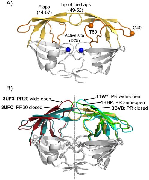
Figure 1.
X-ray structures of HIV-1 PR. A) Structure of PR with flaps in the closed conformation (PDB entry 3BVB), highlighting the flaps (yellow), the hinge regions (orange) and the active site (blue). B) Comparison of the different structural models used in this study, showing the flap conformations of PR in a closed (3BVB, yellow), semi-open (1HHP, green) and wide-open (1TW7, cyan) conformation, as well as the PR20 structures used as reference models for the closed (3UFC, dark cyan) and wide-open (3UF3, red) flap conformations. All models have been superimposed to yield a minimal Cα rmsd for the Core region (comprising residues 10-23, 62-73 and 87-93 of both monomers).

