
Figure 3.
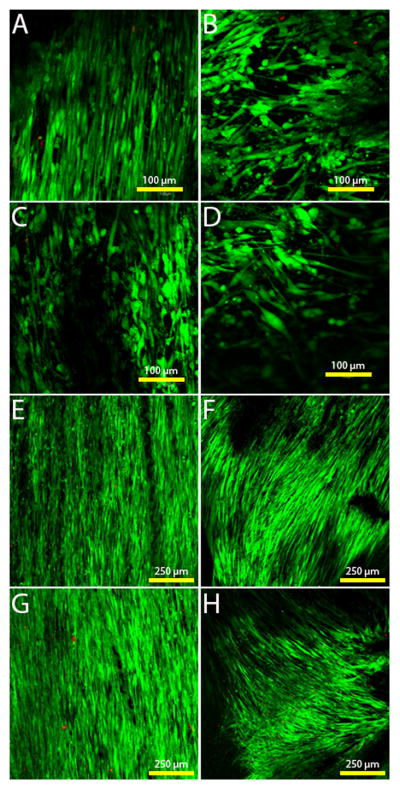
Live/dead staining of the cells on the different scaffold types. Green = viable cells, red = dead cells. A) Aligned scaffold with fibronectin coating on day 3. B) Unaligned scaffold with fibronectin coating on day 3. C) Aligned scaffold with ELP4 coating on day 3. D) Unaligned scaffold with ELP4 coating on day 3. E) Aligned scaffold with fibronectin coating on day 7. F) Unaligned scaffold with fibronectin coating on day 7. G) Aligned scaffold with ELP4 coating on day 7. H) Unaligned scaffold with ELP4 coating on day 7.