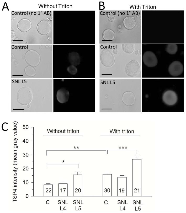
Figure 5.
Increased expression of TSP4 in cultured neurons dissociated from DRGs harvested from nerve-injured rats. (A) Without Triton X-100 treatment, which facilitates antibody penetration into cells, neurons from injured DRGs show elevated surface-bound TSP4. Left-side panels show bright field images. (B) With treatment of Triton, neurons from injured DRGs show increased intracellular TSP4 compared with controls. (C) Summary data. Scale bars = 25μm.*p < 0.05, **p < 0.01, ***p < 0.01.

