Fig. 8.
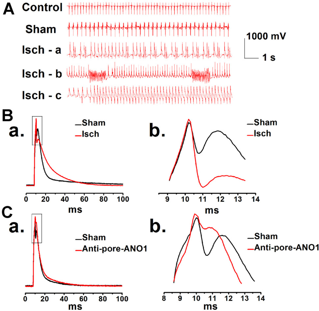
A. Representative ECG recordings in mice during surgeries. Isch-a: an example of ischemia with ventricular premature beats (VE); Isch-b: an example of ischemia with ventricular fibrillation (VF); Isch-c: an example of ischemia with ventricular tachycardia (VT). B. Representative APs were recorded in the mVMs isolated from sham (the black line) and myocardial ischemia (the red line) groups, respectively (B,a). Amplified portion of AP (square boxes in B,a) is shown in panel B,b. C. Representative APs were recorded in the same ventricular myocytes isolated from control mice, before (the black line) or after application of specific pore-targeting anti-ANO1 antibody (the red line) (C,a). C,b. The enlarged portion indicated by square boxes in C,a.

