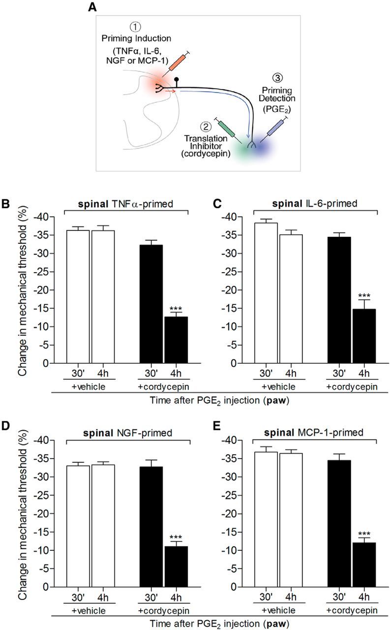
Figure 4.

Hyperalgesic priming at the central terminal of the nociceptor, in the spinal cord, depends on local protein translation. Rats that received spinal intrathecal injection of TNFα (B, 20 ng/μl, 20 μl), IL-6 (C, 1 ng/μl, 20 μl), NGF (D, 150 ng/μl, 20 μl), or MCP-1 (E, 20 ng/μl, 20 μl) 1 week before were tested for hyperalgesic priming of the response to PGE2 (100 ng) injected intradermally into the dorsum of the hindpaw, in the presence or absence of the protein translation inhibitor cordycepin (1 μg, injected at the same site as PGE2 15 min before). Average paw withdrawal thresholds before the intrathecal injections of the priming stimuli and before the injection of PGE2 (1 week later) were as follows: TNFα, 120.6 ± 2.7 and 119.0 ± 2.2 g, respectively, for the vehicle-treated group (t(5) = 0.7734; p = 0.4743, NS) and 118.3 ± 1.4 and 118.3 ± 2.0 g, respectively, for the cordycepin-treated group (t(5) = 0.000; p = 1.000, NS); IL-6, 120.6 ± 1.8 and 120.0 ± 1.2 g, respectively, for the vehicle-treated group (t(5) = 0.3953; p = 0.7089, NS) and 122.6 ± 1.6 and 121.3 ± 2.9 g, respectively, for the cordycepin-treated group (t(5) = 0.0984; p = 0.5160, NS); NGF, 121.6 ± 2.2 and 119.6 ± 1.4 g, respectively, for the vehicle-treated group (t(5) = 1.369; p = 0.2292, NS) and 119.0 ± 1.8 and 118.6 ± 0.8 g, respectively, for the cordycepin-treated group (t(5) = 0.1465; p = 0.8893, NS); MCP-1, 118.6 ± 0.8 and 117.3 ± 1.2 g, respectively, for the vehicle-treated group (t(11) = 0.9834; p = 0.3466, NS) and 120.6 ± 1.0 and 119.6 ± 0.9 g, respectively, for the cordycepin-treated group (t(11) = 0.9199; p = 0.3774, NS); paired Student's t test showed no significant difference between these two values. The nociceptive mechanical paw withdrawal threshold was evaluated 30 min and 4 h after PGE2 injection. Two-way repeated-measures ANOVA followed by Bonferroni post-test showed significant attenuation of PGE2-induced hyperalgesia at the fourth hour after injection in the groups pretreated with cordycepin (***p < 0.001 in all cases, when vehicle- and cordycepin-treated groups are compared at the fourth hour; B, F(1,10) = 126.19; C, F(1,10) = 53.36; D, F(1,10) = 60.63; E, F(1,22) = 63.07; p < 0.0001 for all cases, when vehicle and cordycepin groups are compared), indicating a role of local protein translation in hyperalgesic priming induced by spinal injection of TNFα, IL-6, NGF, or MCP-1 [N = 3 rats (6 paws, B–D) or N = 6 rats (12 paws, E) per group]. A, In the schematic, the red arrow represents the signal triggered by the spinal injection (red syringe) of the priming agents (1, TNFα, IL-6, NGF, and MCP-1) originating in the central terminal of the nociceptor and the subsequent signal (blue arrow) to the peripheral nociceptor terminal (in skin) where hyperalgesic priming is detected by the prolongation of hyperalgesia evoked by PGE2 injected in the paw (2, blue syringe). The green syringe represents the site where cordycepin (or vehicle) was injected.
