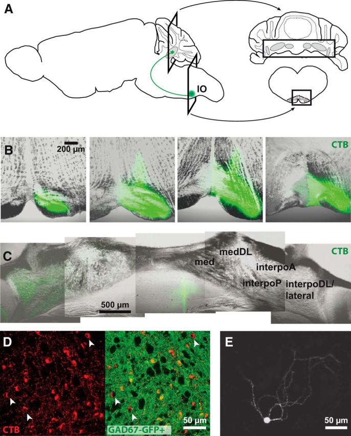
Figure 1.

Retrograde labeling of nucleo-olivary neurons. A, Schematics of the brain (left) showing the CbN (gray) and a nucleo-olivary cell (green) and coronal sections (right) of the cerebellum (top) and brainstem with IO (bottom). Rectangles indicate sections in B and C. B, Confocal images of CTB injections (green) in the IO. Each image from left to right progresses 320 μm rostrocaudally. C, Retrograde labeling in the contralateral anterior and posterior interpositus nuclei (interpoA and interpoP), dorsolateral horn of the interpositus nucleus (interpoDL), and lateral nucleus but not medial nucleus (med) nor its dorsolateral horn (medDL). Montage of confocal images. D, Z-stack of confocal images showing labeled nucleo-olivary cells (red) in GAD67-GFP+ mice (GFP, green) in the lateral nucleus. Arrowheads, GAD67-GFP-negative nucleo-olivary cells. E, Z-stack of confocal images showing a biocytin-filled nucleo-olivary cell with sparsely ramified dendrites. Background is reduced to facilitate visualization of dendrites.
