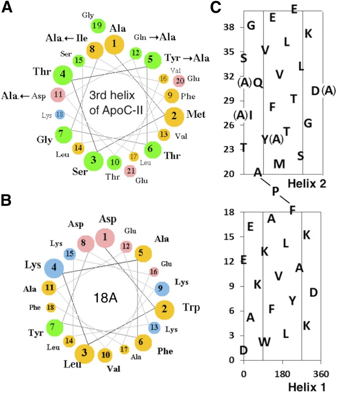
Fig. 1.
Helical wheel and net plots of apoC-II mimetic peptides. (A) Helical wheel plot of the third helix of apoC-II. Arrows show the site of Ala substitutions for the C-II-i peptide. The size of balls and color indicate the degree of charge and hydrophobicity (yellow, nonpolar; green, polar/uncharged; pink, acidic; blue, basic). (B) Helical wheel plot of 18A helix. (C) Helical net plot of C-II-a peptide. The central region indicates the position of the hydrophobic face. Arrows show sites of Ala substitutions for the C-II-i peptide. The first helix (1–18) is based on the 18A helix. The second helix (20–40) is based on the third helix of apoC-II.

