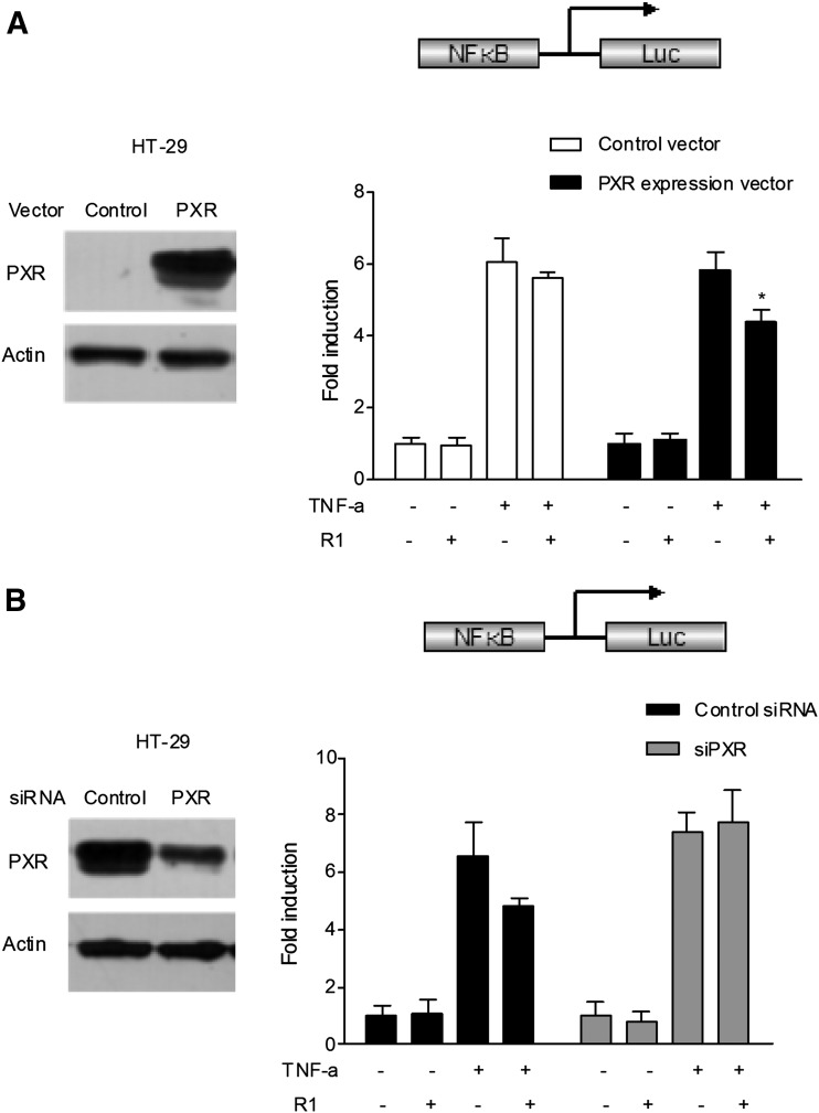
Fig. 4.
Role for PXR in R1-mediated NF-κB luciferase (Luc) inhibition. (A) HT-29 cells were electroporated with the pGL4.32[luc2P/NF-κB-RE/Hygro] reporter combined with pRL-TK and pSG5-hPXR or pSG5-mock. PXR overexpression was verified by Western blot analysis (left). After transfection, the cells were treated with TNF-α (20 ng/ml) alone or in combination with R1 (25 μM) for 24 hours. A standard dual-luciferase activity of the cell lysates was measured, and the results are expressed as the fold induction compared with that of the control cells (designated as 1). The results are presented as the means ± S.D. of three independent experiments. *P < 0.05 versus hPXR-transfected cells treated with TNF-α alone. (B) The human PXR gene was silenced by PXR siRNA transfection in the aforementioned HT-29 cells previously transfected with the pGL4.32[luc2P/NF-κB-RE/Hygro] reporter and the human PXR expression plasmid pSG5-hPXR. The depletion of PXR by siRNA transfection in HT-29 cells was verified using Western blot (left). After transfection, the cells were treated with TNF-α (20 ng/ml) alone or cotreated with R1 (25 μM) for 24 hours. A standard dual luciferase assay of the cell lysates was performed. The results are presented as the means ± S.D. of three independent experiments. *P < 0.05 versus control siRNA transfection samples treated with TNF-α alone.

