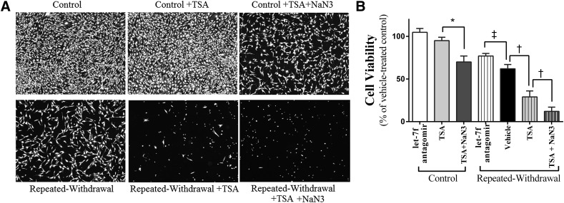
Fig. 6.
Effects of histone acetylation on cell viability. HT22 cells were subjected to an ethanol program consisting of two cycles of ethanol exposure (0 or 100 mM) for 20 hours and withdrawal for 4 hours. TSA (0 or 400 nM) with or without NaN3 (0 or 5 µM), and let-7f antagomir (50 nM) alone were applied during EW phases or the corresponding time period of control cells. At the end of the program, cell viability was assessed using a calcein-AM assay. Microscopic cell populations were assessed using a fluorescence microscope at the objective magnification of 100× (A). Fully confluent, healthy cells with spindle-shaped morphology are shown in control cells. The viability of control cells was not altered by TSA treatment alone but decreased with the combination of TSA with the CcO inhibitor NaN3 (*P < 0.01). Healthy cells with spindle-shaped morphology were much less populated, hardly shown, or did not exist in EW cells treated with vehicle, TSA, or TSA + NaN3, respectfully. Except for control cells treated with TSA or let-7f antagomir, all treatment groups showed statistically significantly lower cell viability (P < 0.01) than the control cells treated with vehicle at dash line (statistical symbols of this difference are omitted for figure clarity); *P < 0.01 versus control cells treated with TSA; †P < 0.005 versus EW cells treated with vehicle or TSA + NaN3; ‡P < 0.01 versus EW cells treated with let-7f antagomir. Depicted values are mean ± S.E.M. for 16 wells/group.

