
Figure 7. Cep126 is involved in cilium formation.
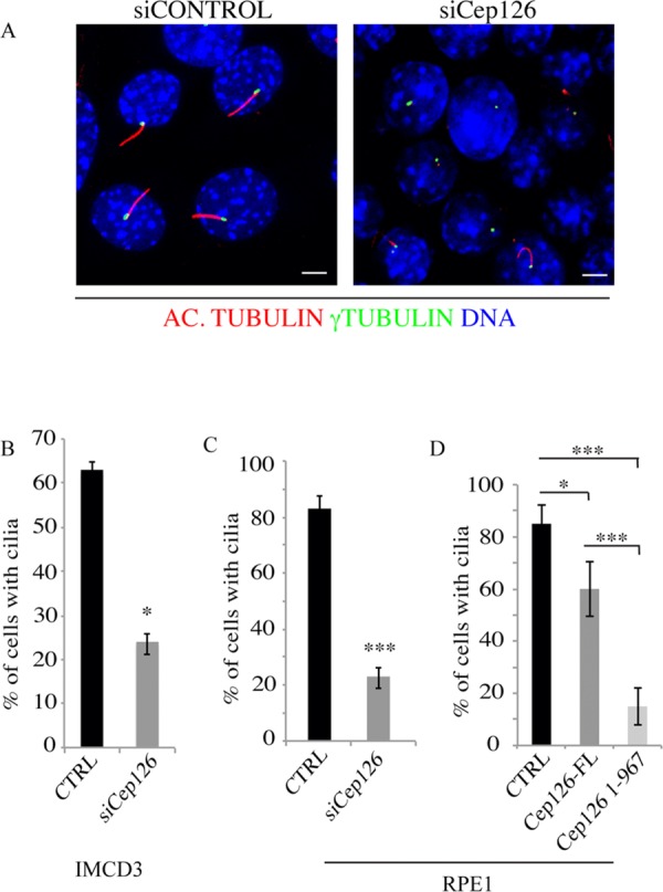
(A) Representative images of IMDC3 cells transfected with non-targeting (siCONTROL) and anti-Cep126 siRNAs (siCEP126), and incubated for 48 h in the absence of serum. The cells were fixed in methanol and stained with an anti-acetylated tubulin antibody (red) and an anti-γ-tubulin antibody (green). The presence of a detectable cilium was monitored by confocal microscopy. (B and C) Quantification of the cells treated as in (A) for the percentages of IMCD3 (B) and hTERT-RPE-1 (C) cells showing a detectable cilium; the reduction of cilium formation was statistically significant. (D) hTERT-RPE-1 cells were transfected with an empty vector as the control (CTRL), and full-length Cep126 (Cep126-FL) and 1–967 Cep126 depletion mutant (Cep126 1–967). The cells were serum starved for 48 h, and fixed and treated for immunofluorescence to monitor cilium formation. Data in (B–D) are means ± SD of three independent experiments; more than 200 cells where counted per experimental condition. Scale bars: 4 µm. *P < 0.001, ***P < 0.0001, using t-test.