Figure 1.
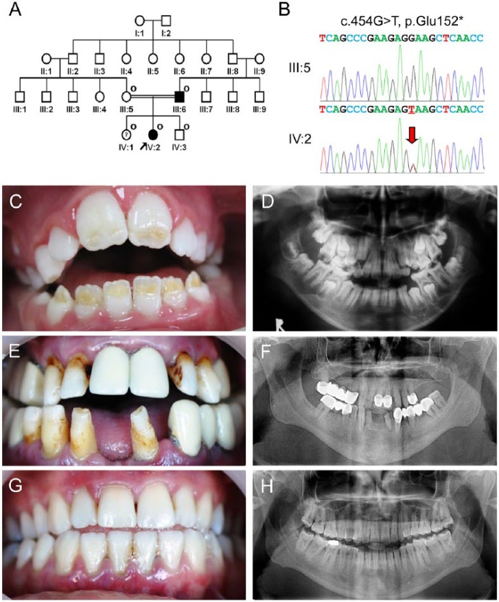
Clinical and mutational analysis of family 1. (A) Pedigree of family 1. The “O” symbol indicates members recruited for this study. The proband is indicated with a black arrow. The question symbol (?) indicates that this individual is clinically normal but has the mutation. (B) Comparison of the ENAM exon 7 sequencing chromatograms for the unaffected family member (III:5) with the wild-type (top) sequence, and the mutated allele in the proband (IV:2) reveals a G-to-T transversion: c.454G>T, p.Glu152*. A red arrow indicates the mutated nucleotide. (C) Frontal clinical photo of the proband. Yellow dentin color can be seen through the hypoplastic enamel located on the incisal half of the permanent anterior teeth. (D) Panoramic radiograph of the proband reveals unusual crown of the developing second permanent molars due to hypoplastic enamel. (E) Frontal clinical photo of the affected father shows the characteristic horizontal hypoplastic grooves. (F) Panoramic radiograph of the affected father shows multiple prosthetics and thin enamel in remaining teeth. (G) Frontal clinical photo of a sister of the proband. Despite having the mutation, her dentition has no enamel defects. (H) Panoramic radiograph of a sister of the proband reveals normal-looking tooth structures.

