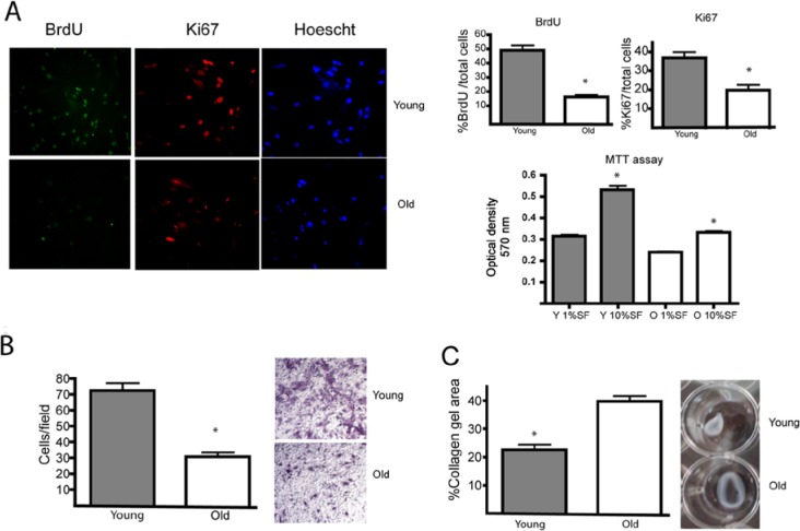
Figure 1.
Cellular responses associated with gingival repair are decelerated in aged HGFs. (A) Young (Y) and old (O) HGFs were incubated with 2 μg/mL BrdU for 24 hrs in the presence of 10% FBS. To evaluate cell proliferation, we stained cells for BrdU and Ki67. Magnification bar equals 10 μm. Graph indicates average and standard error of positive nuclei for BrdU (+) and Ki67 (+) cells normalized against total cells stained with Hoechst. MTT assay: Graph indicates average and standard error of OD at 570 nm. Asterisks indicate statistically significant differences between young and old fibroblasts. (B) Young and old HGFs were placed in the upper compartment of a transwell chamber. In the lower chamber, 10% FBS was added. Migration was allowed to occur for 16 hrs. Quantification of migration assay is expressed as the average number of migrating cells detected in the lower side of the filter. Young fibroblasts were more migratory than old fibroblasts (p < .001). Magnification bar equals 50 μm. (C) Young and old HGFs were cultured within collagen gels (1 mg/mL) and exposed to 10% FBS. Collagen gel areas are represented as average ± standard error. Young HGFs were more contractile than old HGFs (p < .001). Bars indicate standard error. All assays were performed in quadruplicate. All assays were performed in cell lines derived from 4 different donors.

