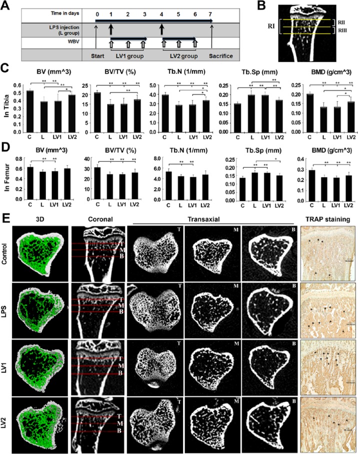
Figure 2.
Immediate exposure to whole body vibration (WBV) reduces lipopolysaccharide (LPS)–mediated trabecular bone loss of the tibia. (A) Schematic representation of experimental protocols. The mice were divided into a control group (C), LPS-treated group (L; 5 mg/kg), and LPS + WBV group (LV). Mice of the LV group were exposed to WBV (45 Hz, 0.4 g) the day after the first LPS injection (LV1) or the day after the second injection (LV2) for 10 min/d for 3 d. All animals were sacrificed 7 d after the first injection of LPS. (B) According to micro–computed tomography (micro-CT) analysis, the 1-mm-long area under the growth plate of the metaphyseal area (region 1) of each group was divided into the proximal 0.5-mm portion (region 2) and the distal 0.5-mm portion (region 3) to identify the most sensitive region to LPS treatment. RI, RII, and RIII indicate regions 1, 2, and 3, respectively. (C) Histomorphometric parameters obtained at the end of the experiment include bone volume/tissue volume (BV/TV), trabecular number (Tb.N), trabecular separation (Tb.Sp), and bone mineral density (BMD) of region 1. Values are expressed as mean ± SD. * p < .05 and ** p < .01 indicate significant differences between the 2 groups. (D) BV and BMD of the femur in RI (defined as for the tibia). (E) Micro-CT images of coronal sections and transaxial sections are presented at the top, middle, and bottom portions of RI. Three-dimensional reconstructions of RIII support quantifiable changes in both density and architecture of the bone. Representative images of immunohistochemical staining for TRAP expression are presented to detect osteoclasts. Marked positions with asterisks correspond to cells. Scale bar indicates 100 µm.

