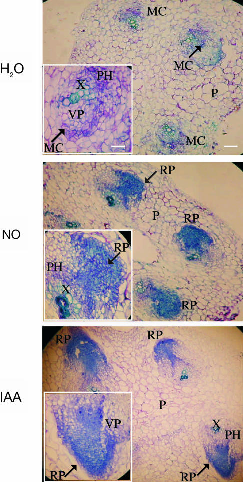
Figure 1.
Primordia formation during adventitious root development in cucumber explants. Primary roots were removed and the explants were treated with water or 10 μm of the NO-donor SNP or 10 μm IAA for 3 d. Hypocotyl transverse sections were stained with toluidine blue and photographed using a digital camera attached to the microscope. For photographs and insets, magnifications of ×100 (bar indicates 0.2 mm) and ×400 (bar indicates 0.05 mm) were respectively used. MC, meristematic cells; P, parenchyma; PH, phloem; RP, root primordium; VP, vascular parenchyma; X, xylem. Arrows indicate the MC or RP magnified in the insets.

