
Figure 2.
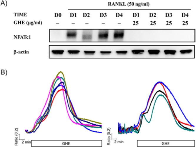
GHE provokes a transient [Ca2+]i elevation, resulting in suppression of RANKL-mediated NFATc1 expression. (A) Isolated BMMs were treated with RANKL (50 ng/mL, D0 presents DW treatment instead of RANKL) in the presence or absence of GHE (25 μg/mL) and cultured for indicated times (0 ~ 4 days). Following incubation, whole-cell lysates were collected and used for the evaluation of total NFATc1 expression. (B) BMMs seeded onto cover glass were maintained for 2 days with (right panel) or without RANKL stimulation (left panel). After incubation, intracellular Ca2+ mobilization in a single cell was measured as described in Materials & Methods. Cells were perfused with HEPES buffer, and then GHE (25 μg/mL) diluted in HEPES buffer was acutely treated until the end of the experiment. Each trace presents cytosolic Ca2+ mobilization in each cell.