
Figure 3.
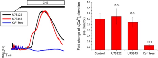
GHE-mediated [Ca2+]i elevation is dependent on an influx of extracellular Ca2+, but not on PLC activation. (Left panel) BMMs seeded on cover glass were incubated in the presence of RANKL (50 ng/mL) for 2 days and then used for intracellular Ca2+ measurement. Prior to GHE, cells were acutely treated with U73122 (10 μM, PLC inhibitor, black line), U73343 (10 μM, inactive control for U73122, red line), and EGTA (1 mM) diluted in HEPES buffer (without CaCl2, blue line). In the presence of U compounds or EGTA, GHE was added and maintained until the end of each experiment. The columns on the right show the maximal [Ca2+]i levels provoked by GHE. Data are presented as mean ± SD of at least 3 independent experiments. ***p < .001 vs. the control group, which was treated with only GHE in the absence of U compounds and EGTA.