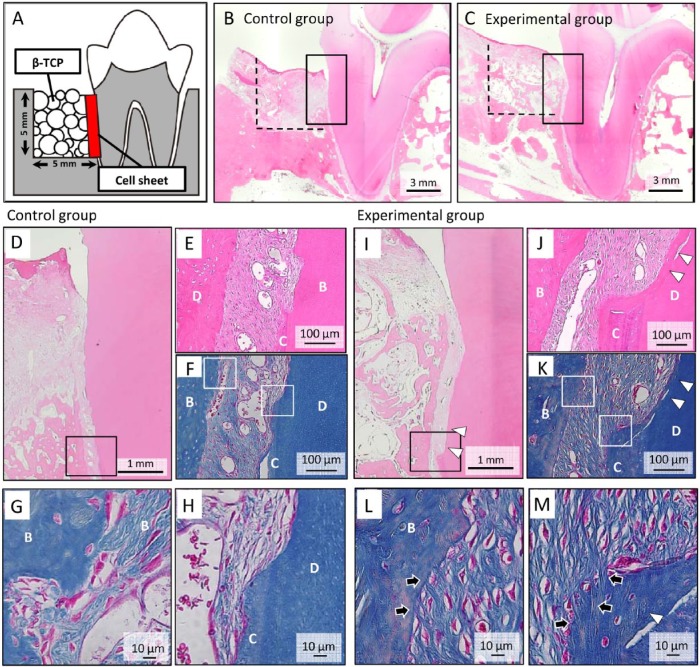
Figure 4.
Periodontal tissue regeneration with autologous transplantation of dog dental socket–derived cells (dDSCs). (A) Schematic illustration of a one-wall periodontal defect in the dog mandible. (B-E) After 3-layered dDSC sheets with polyglycolic acid were transplanted onto the surface of the tooth root, β-TCP was implanted into the one-wall bone defect. A histologic analysis was performed 2 mo after transplantation (B, D-H: control group; C, I-M: experimental group). (D, I) Higher magnification of the squared areas in Figure 4B and 4C, respectively. A greater amount of newly formed bone was observed in dDSC-transplanted group than that in the control group. The squares indicate the areas shown in Figure 4E (hematoxylin and eosin staining) and 4F (Azan staining) or 4J (hematoxylin and eosin staining) and 4K (Azan staining) at a higher magnification. (H, G) Higher magnification of Figure 4F. (L, M) Higher magnification of Figure 4K. Sharpey’s fiber-like tissue (arrow) connecting the newly formed bone and cementum-like tissue (arrowhead) was observed in the experimental group. B, bone; C, cementum; D, dentin.

