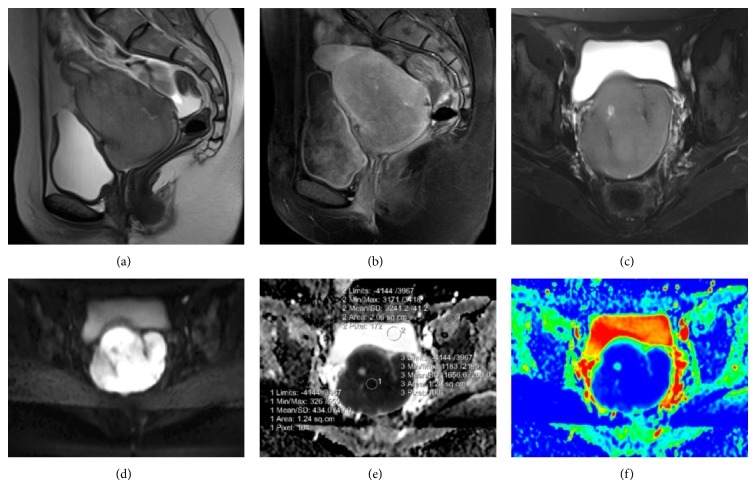
Figure 1.
(a) Sagittal T2-weighted imaging shows a slightly hyperintense mass confined to the cervix. Free intraperitoneal fluid is evident. (b) Sagittal postcontrast T1-weighted imaging displays heterogeneous enhancement of the mass. (c) Cystic area is seen on axial T2 fat suppressed image. (d) Axial TRACE images reveal prominent diffusion restriction in the mass lesion. (e, f) Corresponding ADC and coloured ADC maps confirm high cellularity.

