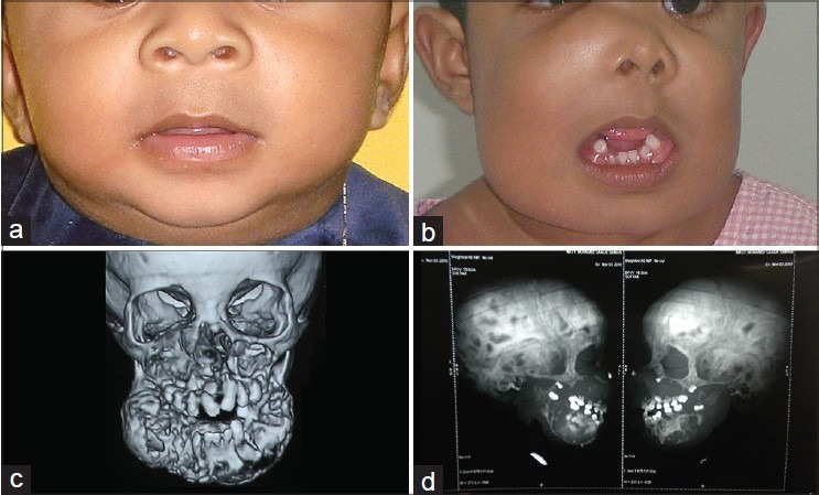
Figure 1.

(a) Patient preoperative view at the age of 7 months. (b) Preoperative view at the age of 18 months with the right side facial swelling obliterating the right nasolabial fold, elevating the right lower eyelid. Note the mandibular swelling which is extending from angle to angle. (c and d) Preoperative computed tomography showing the well-defined, multiple solitary radiolucencies with radiopaque sclerotic margin, note the welldefined mandibular radiolucency with radiopaque border sparing bilateral condyle ramal unit and dentoalveolar segment of the anterior mandible
