Figure 6.

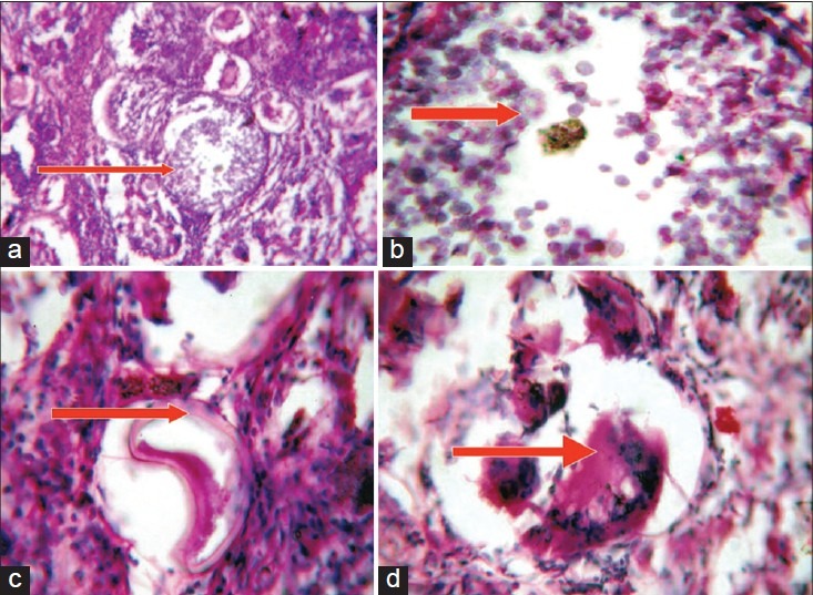
Photomicrographs [×10 (a, b), ×40 (c, d) H and E], showing mature sporangium (a) surrounded by dense stroma infiltrated by inflammatory cells, endospores (b), collapsed cyst (c) and foreign body giant cell reaction (d)

Photomicrographs [×10 (a, b), ×40 (c, d) H and E], showing mature sporangium (a) surrounded by dense stroma infiltrated by inflammatory cells, endospores (b), collapsed cyst (c) and foreign body giant cell reaction (d)