
Figure 1.
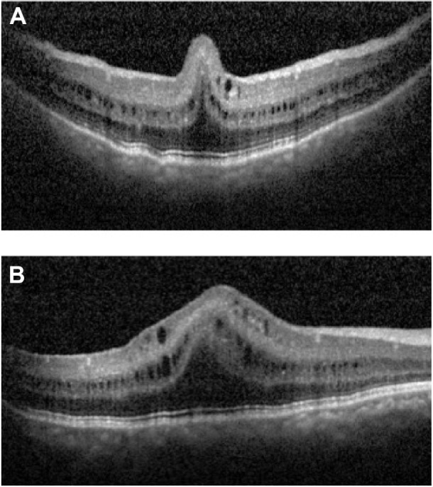
SD-OCT of the macula in the right eye at presentation.
Notes: Both the vertical (A) and the horizontal (B) scan show a PMF with cyst-like cavities in the inner nuclear and ganglion cell layers. The vertical scan (A) shows the high, inverted U pattern of the PMF, and the horizontal scan (B) passing through the PMF clearly demonstrates the cystic cavities extending beyond the PMF. Note the preservation of the stratification of the retinal layers throughout the macula and the absence of involvement of the majority of the outer retina by the PMF.
Abbreviations: SD-OCT, spectral-domain optical coherence tomography; PMF, papillomacular fold.