Fig. 3.

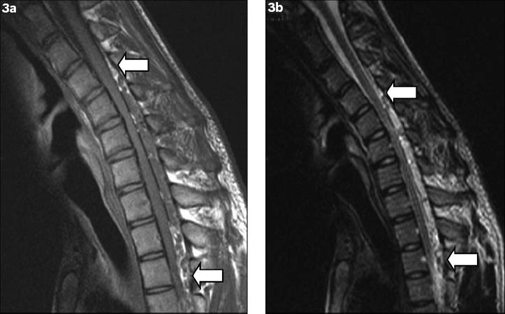
Flexion (a) T1-W and (b) T2-W sagittal MR images show anterior displacement of the posterior wall of the cervical dural canal from levels C4 to T3 (arrows), with flattening of the cord and complete effacement of the anterior thecal sac.

Flexion (a) T1-W and (b) T2-W sagittal MR images show anterior displacement of the posterior wall of the cervical dural canal from levels C4 to T3 (arrows), with flattening of the cord and complete effacement of the anterior thecal sac.