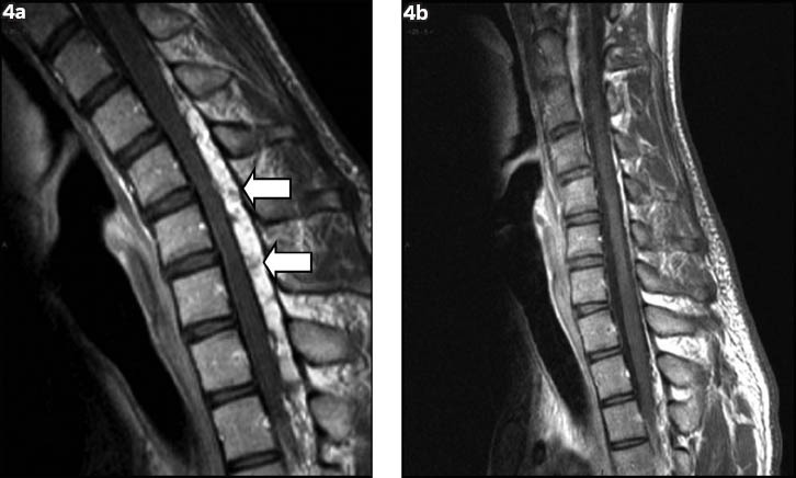
Fig. 4.

Dynamic T1-W postcontrast sagittal flexion MR images show (a) a homogeneously contrast-enhancing posterior epidural mass with multiple small signal voids within (arrows); and (b) disappearance of the epidural mass when the neck returns to a neutral position.
