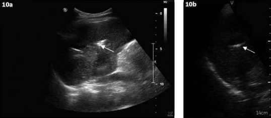
Fig. 10.

Intrahepatic stones. (a) Image from the standard ultrasound machine shows linear echogenicity with posterior shadowing (arrow) within the liver parenchyma, which is suggestive of intrahepatic stones. (b) Image from the pocket-sized ultrasound machine shows similar-looking linear echogenicity with posterior shadowing (arrow).
