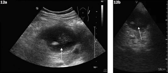
Fig. 12.

Intra-abdominal collection. (a) Image from the standard ultrasound machine shows a loculated hypoechoic area with internal echogenicity (arrow), which is suggestive of collection. (b) Image from the pocket-sized ultrasound machine shows a similar-looking hypoechoic collection (arrow).
