Figure 6. .
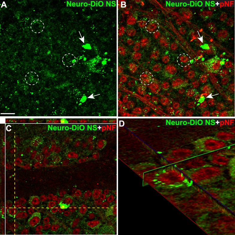
RGCs take up Neuro-DiO released from 50 nm NS. Confocal micrographs of whole mount retina 1 week after intravitreal injection of Neuro-DiO loaded NS. (A) Neuro-DiO (green) was released from NS and deposited on the retinal surface (arrows). (B) Uptake of Neuro-DiO by phosphorylated neurofilament-positive RGCs (pNF; red) was observed (dotted circles). (C) Confocal micrograph and orthogonal projections showing Neuro-DiO surrounding pNF-positive RGC somas. The yellow dotted lines indicate the position of the orthogonal views. (D) An orthogonal view rotated about the Z-axis shows Neuro-DiO deposits surrounding a pNF-positive RGC. Scale bar: 10 μm.

