
Figure 4.
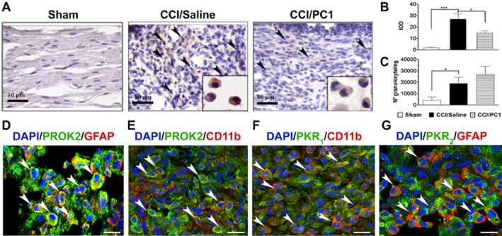
Representative images of sciatic nerve in the immediate proximity of the injury. (A) Immunohistochemical staining of ipsilateral sciatic nerve, on day 0 after CCI, from sham, CCI/saline and CCI/PC1 mice with anti-PROK2 antibody and haematoxylin. Scale bar = 30 µm. Arrowheads indicate the infiltrating cells expressing the PROK2 protein. A sustained infiltration of PROK2-positive cells was evident 10 days after CCI. PC1 treatment significantly reduced the PROK2 immunoreactivity (brown colour) in the cytoplasm of these cells (inset) as demonstrated in (B) by quantitative analysis of PROK2 signal computed as integrated optical density for arbitrary areas (six sections per animal, six animals). (C) Number of infiltrating neutrophils evaluated as MPO activity mg−1 tissue. Data are means ± SEM of four to six animals. *P < 0.05; ***P < 0.001 CCI/saline versus sham mice; °P < 0.05 CCI/PC1 versus CCI/saline mice; one-way anova, followed by Tukey's test for multiple comparisons. (D and E) Immunofluorescence double staining showing colocalization (yellow, arrowheads) of PROK2 (green) with GFAP (Schwann cell marker, red) and CD11b (macrophage marker, red) in the immediate proximity of the injury in the sciatic nerve of CCI/saline mice. (F and G) Representative images showing the localization (arrowheads) of the receptor PKR1 (green) in CD11b-positive macrophages (red) and of the receptor PKR2 (green) in GFAP-positive Schwann cells (red) in the immediate proximity of the injury of the sciatic nerve in CCI/saline mice. Cell nuclei were counterstained with DAPI (blue fluorescence). Scale bar, 20 µm.