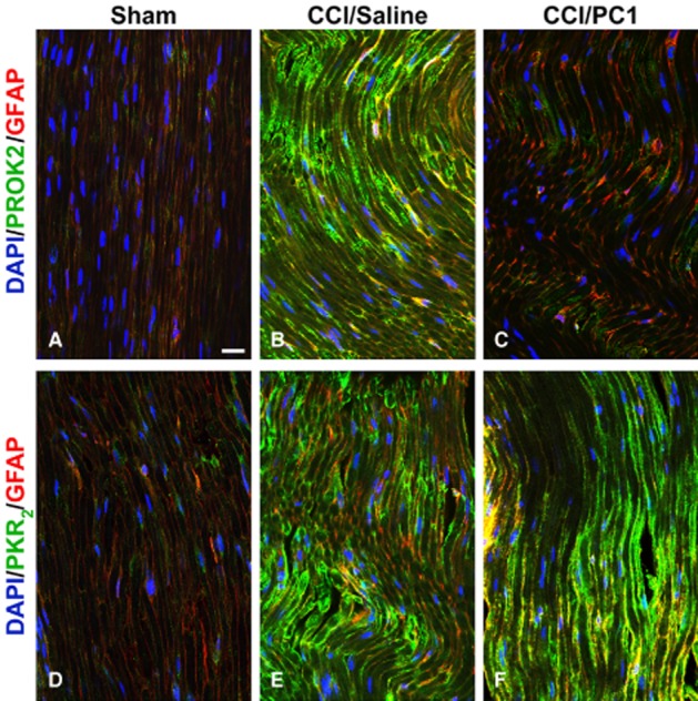
Figure 5.

Representative images of CCI-induced up-regulation of PROK2 and PKR2 in the longitudinally sliced sciatic nerve proximal to the lesion. PROK2 immunofluorescence was never found in uninjured nerve (A). Only a very faint PKR2 signal was evident in the non-activated Schwann cells (GFAP-positive cells, red) (D). A dramatic increase of PROK2 and PKR2 signal (B and E, green) in fibres and in GFAP-positive structures was evident 10 days after nerve ligation. PC1 treatment prevented the injury-induced PROK2 up-regulation (C) but was ineffective against PKR2 up-regulation (F). Cell nuclei were counterstained with DAPI (blue fluorescence). Scale bar: 20 µm.
