
Figure 8.
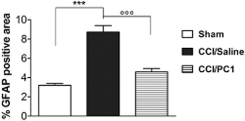
Quantification of the area occupied by the astrocyte marker GFAP in 300 µm2 area, intersecting laminae I and II of cross-sectional spinal cord from sham, CCI/saline and CCI/PC1 mice (five sections per animal, three animals per group). PC1 treatment significantly reduced the CCI-induced astrocytosis and microgliosis. Data are mean ± SEM of four to six animals. ***P < 0.001 CCI/saline versus sham mice; °°°P < 0.001 CCI/PC1 versus CCI/saline mice; one-way anova, followed by Tukey's test for multiple comparisons.