
Figure 9.
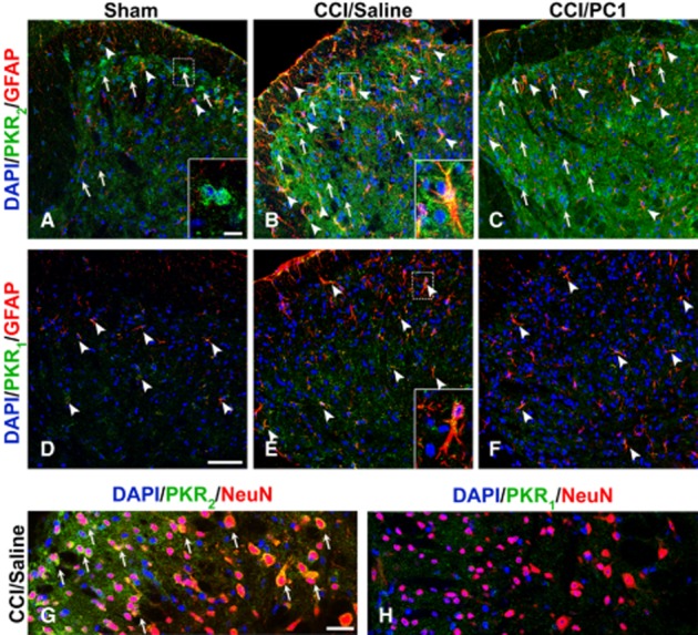
Representative images showing PKR2 and PKR1 localization in the mouse L4–L5 spinal cord dorsal horns from 10 days sham (A, D), CCI/saline (B, E) and CCI/PC1 (C, F) mice. PKR2 immunofluorescence (green) is clearly evident in sham animals, localized in some neuronal cells (a, arrows and inset), and in some astrocytes (A, arrowheads). Ten days after CCI (B), PKR2 positive neuronal cell bodies were more evident also in deeper layers of the dorsal horn (B, arrow) as demonstrated by colocalization with the neuronal marker NeuN (G). The localization of PKR2 in activated astrocytes is demonstrated by the double staining of PKR2 (green) with the astrocytes marker GFAP (B, arrowheads and inset). The diffuse punctuate pattern PKR2 signal appeared clearly increased. PC1 treatment did not modify the PKR2 immunofluorescence intensity. In the spinal cord, the PKR1 signal was very faint and was not affected by nerve injury nor by PC1 treatment. PKR1 immunoreactivity was clearly evident in GFAP-positive resting and activated astrocytes (D, E) and was not modified by PC1 treatment. We never detected PKR1 signal in NeuN-positive cells. Cell nuclei were counterstained with DAPI (blue fluorescence). Scale bar, 50 µm in A to F; 30 µm in G, H and 10 µm insets.